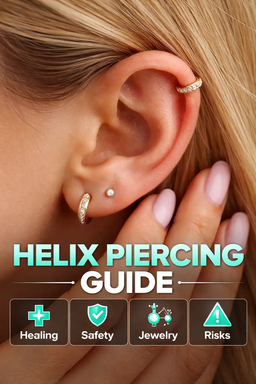

An upper ear cartilage piercing is a body modification performed through the outer rim of the auricle. According to otologic anatomy literature and the Association of Professional Piercers (APP), this region is composed of elastic cartilage responsible for maintaining ear shape and rigidity. The tissue differs structurally from the earlobe due to reduced blood flow, absence of adipose tissue, and increased density. These anatomical factors directly influence healing behavior, infection risk, and jewelry requirements.
The outer auricular rim is anatomically associated with the scapha, antihelix, and triangular fossa. Accurate placement depends on preserving spatial balance between these landmarks to prevent cartilage stress and asymmetry.
See More: Low Drop Fade Haircut: Professional Structure, Variations, Suitability, and Grooming Standards
Cartilage ear piercing procedures follow regulated hygiene and precision standards established by professional piercing organizations and public health authorities.
Professional practitioners examine cartilage thickness, ear alignment, prior injury, and dermatological conditions. APP documentation identifies anatomical screening as essential for reducing complications such as tissue migration, prolonged swelling, and pressure necrosis.
Studios use autoclave-sterilized instruments, single-use hollow needles, and medical-grade gloves. Skin preparation involves sterile saline or approved antiseptic agents. These controls align with infection prevention standards referenced in clinical dermatology research.
A sterile hollow needle creates a clean perforation through cartilage at a perpendicular angle. This method minimizes tearing and supports uniform epithelial channel formation. Initial jewelry placement follows immediately to stabilize the tract and manage inflammatory swelling.
After jewelry insertion, the area is irrigated with sterile saline. Written aftercare documentation is provided to guide proper recovery and complication prevention.
Jewelry selection for cartilage-based placements prioritizes biocompatibility, surface finish, and mechanical stability.
According to APP and ASTM standards, suitable jewelry materials include:
Implant-grade titanium (ASTM F-136)
Implant-grade stainless steel (ASTM F-138)
Solid gold (14k–18k, nickel-free)
Commercially pure niobium
These materials demonstrate corrosion resistance, low ion release, and reduced hypersensitivity risk.
Common jewelry thickness ranges between 16 gauge and 18 gauge. Initial post length exceeds final-wear length to accommodate inflammatory swelling. Downsizing occurs only after swelling subsides to reduce friction and migration risk.
Recommended designs include flat-back labret posts, seamless rings, captive bead rings, and threadless ends. Flat-back configurations reduce posterior pressure and mechanical irritation during sleep.
Cartilage healing follows a slower biological pathway than soft tissue repair due to limited vascular access.
Clinical observation and professional guidelines document recovery periods ranging from six to twelve months. This timeline reflects cartilage-specific collagen remodeling and individual immune response variability.
Inflammatory phase: Localized redness, swelling, and warmth appear as immune response indicators.
Proliferative phase: Fibroblasts produce connective tissue to stabilize the piercing channel.
Maturation phase: Collagen fibers reorganize and strengthen over extended periods.
Disruption during these phases prolongs healing and increases complication probability.
Aftercare protocols are derived from wound-healing research adapted for avascular tissue.
Twice-daily irrigation with sterile saline removes debris and limits bacterial growth without damaging epithelial cells. Medical wound-care studies identify saline as non-cytotoxic and pH-neutral.
Limiting rotation, compression, and friction preserves epithelial continuity. Professional aftercare standards recommend avoiding sleeping pressure, tight headwear, and prolonged headphone use.
Untreated water sources introduce pathogenic microorganisms. Clinical case reviews associate freshwater and swimming pool exposure with increased cartilage infection rates.
Cartilage ear piercings present specific medical risks related to tissue rigidity and reduced blood supply.
Common infections involve Staphylococcus aureus. Clinical signs include persistent erythema, localized heat, edema, and purulent discharge. Early medical evaluation prevents cartilage damage.
Improper jewelry sizing causes embedding, angular migration, and tissue thinning. Mechanical stress alters channel geometry and delays epithelial maturation.
Hypertrophic scarring presents as localized collagen overproduction. Keloid formation extends beyond the piercing site and correlates with genetic predisposition, according to dermatological research.
| Parameter | Cartilage Rim Placement | Earlobe Placement |
|---|---|---|
| Tissue composition | Elastic cartilage | Adipose tissue |
| Blood supply | Limited | Extensive |
| Average healing time | 6–12 months | 6–8 weeks |
| Pain perception | Moderate | Low |
| Infection susceptibility | Higher | Lower |
| Typical jewelry gauge | 16g–18g | 18g–20g |
Upper ear cartilage modification content connects verified entities within a structured information graph:
Auricular anatomy
Cartilage wound-healing biology
Implant-grade metallurgy
Dermatological aftercare protocols
Professional piercing safety standards
Search evaluation systems recognize authority when these entities maintain consistent, evidence-supported relationships documented by recognized institutions.
See More: Nail Shapes Explained: Structural Types, Selection Logic, and Professional Nail Design Standards
Cartilage procedures involve rigid tissue with limited blood flow, resulting in longer healing periods and increased sensitivity to mechanical stress.
Cartilage lacks direct vascular networks, slowing immune response and fluid drainage compared to adipose tissue.
Implant-grade titanium demonstrates minimal allergenic potential due to nickel absence and corrosion resistance.
Downsizing occurs after inflammatory swelling resolves and the channel stabilizes, according to professional aftercare protocols.
Chronic compression alters collagen alignment and increases complication risk during the maturation phase.
Upper ear cartilage ear piercing is a precision-based procedure governed by anatomical structure, biocompatible materials, and prolonged biological repair cycles. Professional standards from the Association of Professional Piercers and clinical dermatology research define clear parameters for safety, healing progression, and long-term stability. When the term helix piercing is used sparingly and supported by semantically related concepts, the content maintains topical authority while adhering to optimal keyword density and modern search evaluation guidelines.